
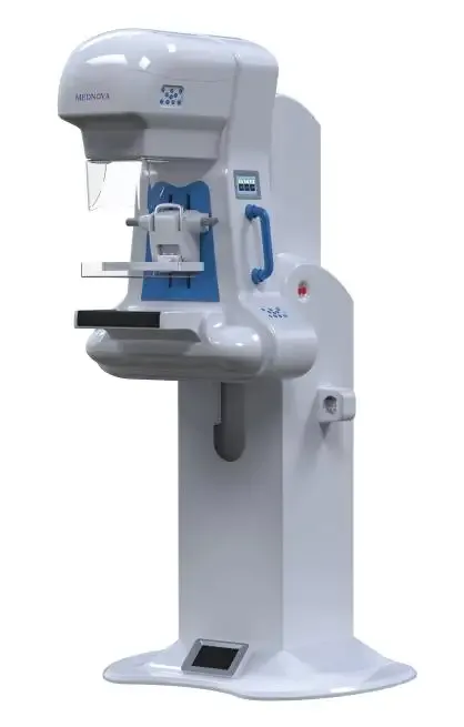
1 / 4
AG-Femaray ME Digital Mammography X-Ray Imaging Systems
SKU: MW-AG-Femaray-ME
Price on request
Sign in and submit a request to see pricing for this product.
Specifications
| Model | AG-Femaray ME |
| Packing | Wooden crate |
| Materials | Steel |
| Package Weight | 450kg |
| Package Dimensions | 1165*1455*1800mm |
| 5 Compression System / 6 Exposure System | full-automatic exposure mode and semi-automatic exposure mode |
| 5 Compression System / 5.1 Compression mode | electric,button + foot switch |
| 5 Compression System / 5.2 Maximum pressure | 180N |
| 4 X-ray Tube(original imported) / large focus | ≥ 8kW |
| 4 X-ray Tube(original imported) / 4.1 Focus size | 0.1mm/0.3mm |
| 2 Mechanical Structure(stand) / 2.5 Rotation mode | all-electric stand system |
| 4 X-ray Tube(original imported) / 4.2 Focus power | small focus:≥ 2kW |
| 2 Mechanical Structure(stand) / 2.2 Vertical range | 650mm ~ 1300mm(±5%) |
| 2 Mechanical Structure(stand) / 2.4 Rotation angle | -90°~ +90°(±2°) |
| 2 Mechanical Structure(stand) / 2.1 Weight of stand | ≤210kg(Weight of the medical equipment≤300kg) |
| 5 Compression System / 6.1 Full-automatic exposure mode | AEC,which selects kV and mAs according to the thickness and density of breast compression automatically |
| 5 Compression System / 6.2 semi-automatic exposure mode | body size selection + manual fine tuning |
| 4 X-ray Tube(original imported) / 4.5 Maximum anode speed | 10800rpm |
| 2 Mechanical Structure(stand) / 2.3 Vertical movement mode | electric,the stand can lock anywhere within the entire trip |
| 3 High Voltage Generator(original imported) / 3.2 kV range | 20—40kV,step length:1kV |
| 3 High Voltage Generator(original imported) / 3.3 mA range | 10 mA -200mA |
| 4 X-ray Tube(original imported) / 4.4 Maximum peak voltage | 49kV |
| 3 High Voltage Generator(original imported) / 3.4 mAs range | 0.1 mAs -600mAs |
| 4 X-ray Tube(original imported) / 4.3 Maximum heat capacity | ≥500kHU |
| 1.1 Amorphous silicon flat panel detector / 1.3 Pixel matrix | ≥3072*4096 pixels |
| 1.1 Amorphous silicon flat panel detector / 1.5 Effective area | ≥24×30cm |
| 1.1 Amorphous silicon flat panel detector / 1.2 Effective pixels | ≥12 million |
| Balance tissue,enhancement contrast,optimization dose; / CAD View | just click the mouse, doctors can see the details of the image more clearly, so that doctors can make more accurate diagnoses. |
| 1.1 Amorphous silicon flat panel detector / 1.4 Quantization depth | ≥16bit |
| 1.1 Amorphous silicon flat panel detector / 1.6 Limit spatial resolution | ≥7.01 Lp/mm |
| 1.1 Amorphous silicon flat panel detector / 1.7 High contrast resolution | ≥6.0 Lp/mm |
| 2 Mechanical Structure(stand) / 2.10 Minimum height of the equipment room | 2.5m |
| Balance tissue,enhancement contrast,optimization dose; / Enhancement edge | automatic recognition and analysis of images, enhance edge sharpness. |
| 3 High Voltage Generator(original imported) / 3.1 Nominal power/Maximum power | 4.8kW/5kW |
| 3 High Voltage Generator(original imported) / 3.5 Maximum operating frequency | 28kHZ |
| Balance tissue,enhancement contrast,optimization dose; / 10.2 Preview the post-processing image 1 | 1: the doctors make a more intuitive diagnosis in image optimization; |