
1 / 2
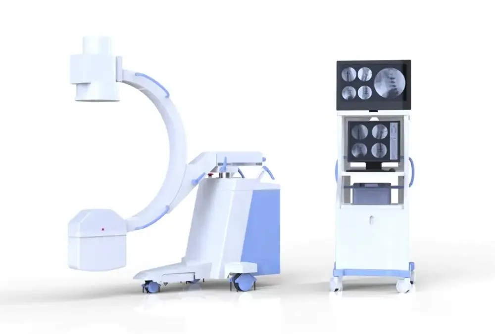
High-Frequency Mobile Digital C-arm System AG-D112B
SKU: MW-AG-D112B
Key features: High-quality combined high-frequency high voltage X-ray generator reduces x-ray dose to the body; With the function of kV, mA automatically tracks (ABS) in fluoroscopy to make the image brightness and clearness; Real-time continuous Pulse Fluoroscopy with a low dose, but meet operation demand with clearness image; Human graphical LCD touch screen, intelligent and fast operation.
Price on request
Sign in and submit a request to see pricing for this product.
Specifications
| GW | 575kg |
| NW | 440kg, |
| Item | Quantity |
| Model | AG-D112B |
| Packing size | 2440*1100*1420mm, |
| Hand controller | 1 |
| I. Usage / Surgery | Removing foreign body, implanting pace maker, partial radiography, local photography, and other surgeries |
| 19’’ LCD monitor | 1 |
| 24’’ LCD monitor | 1 |
| Video System / Monitor | 19’’ LCD monitor and 24’’ LCD monitor |
| Imported mini groove grid | 1 |
| The main frame of the C-arm | 1 |
| Red light cross localization | 1 |
| Structural performance / C-arm | Electric Ascending & Descending of pillar movement:400mm Forward and Backward Movement: 200mm Revolution around Horizontal Axis: ±180° Revolution around Vertical Axis: ±15° Slip-on orbit: 120°(+90°~ -30°) SID: 1000mm Opening: 760mm Arc depth: 670mm |
| Video System / CCD Video camera | Medical Mega-pixels digital CCD camera |
| Video System / Image Intensifier | TOSHIBA 9" image intensifier |
| Electrically adjustable collimator | 1 |
| Video System / Work Station Software | Storage without damage, multi-images display, image W/L real-time adjustment, grayscale switch, region of interest balance, GAMMA correction, flip, noise reduction, intensifying, smoothing, sharpening, compression, zoom in, measurement, remark, picture-text typeset print, expert template, DICOM image transmit, DICOM image print, cine loop, image imprinting, Worklist registration, etc. |
| Fluoroscopy/ Photography / Photography | Tube Voltage:40 kV-120 kV mA:25-100mA mAs:1-180 mAs |
| Medical Mega-pixels digital CCD camera | 1 |
| Structural performance / Directive wheel | Direction wheel 360°rotation, main wheel ±90°rotation |
| Fluoroscopy/ Photography / Plate holder Size | 200mm×250mm(8″×10″)or 250mm×300mm(10″×12″) |
| Fluoroscopy/ Photography / Manual Fluoroscopy | Tube Voltage:40kV~120kV(continuous adjustment) Tube Current:0.3mA~4mA(continuous adjustment) |
| Fluoroscopy/ Photography / Pulsed Fluoroscopy | Tube Voltage:40kV~120kV(continuous adjustment) Tube Current:0.3mA~30mA (continuous adjustment,Intelligent control of pulse frequency) |
| Digital Acquisition and Processing workstation | 1 |
| Toshiba 9’’ three fields image intensifier | 1 |
| Fluoroscopy/ Photography / Automatic Fluoroscopy | Tube Voltage: 40kV~120kV(automatic adjustment) Tube Current:0.3mA~4mA(automatic adjustment) |
| X-ray generator and high-frequency inverter power supply | 1 |
| X-ray Tube Special for High Frequency / Anode Thermal Capacity | 35kJ |
| X-ray Tube Special for High Frequency / Fixed anode Dual-focus | 0.6/1.8mm |
| Fluoroscopy/ Photography / High-Frequency Inverter Power Supply | Output Power:5kW Inverter Frequency:40kHz |