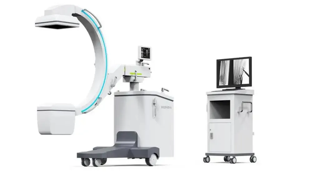
Mobile C-Arm X-Ray Machine AG-CStar_S15.6KW (Dual-mode)
SKU: MW-AG-CStar_S15.6KW-(Dual-mode)
CStar-S1(without wifi) (5.6kW,125kV, 0.6/1.2mm,81kHU, 21cm*21cm) The AG-CStar_S1 mobile c-arm x-ray machine is used to take images of the spine, bones, and soft tissues. It can also be used to guide procedures such as surgery or biopsies. In some cases, mobile c-arm x-ray machines may also be used to take images of the heart or blood vessels. Mobile c-arm x-ray machines use fluoroscopic dyes to produce detailed, real-time X-ray imaging for many different diagnostic and interventional procedures.
Price on request
Sign in and submit a request to see pricing for this product.
3 Flat panel detector / 3.5 Dynamic adjustment range
≥16bit
3 Flat panel detector / 3.1 Dynamic plate detector type
Amorphous silicon CsI flat panel detector
2 X-ray control system / 2.5 Maximum photographic current
80mA
4 C arm Stand system / 4.6 The distance from the focus to the screen
≥ 1000mm
1.1 High-frequency inverter power supply and combined tube / 1.2 Power
5.6kW
1.1 High-frequency inverter power supply and combined tube / 1.3 Focus size
0.6mm ( small focus )/1.2mm ( big focus )
1.1 High-frequency inverter power supply and combined tube / 1.6 Inverter frequency
≥200kHz
1.1 High-frequency inverter power supply and combined tube / 1.5 Anode heat capacity
60kJ(81kHU)
1.1 High-frequency inverter power supply and combined tube / 1.7 Continuous perspective
with manual and automatic perspective modes, the perspective current is 0.2~10mA
1.1 High-frequency inverter power supply and combined tube / 1.4 Heat capacity of tube cover
480kJ ( 648kHU )
Product details
CStar-S1(without wifi) (5.6kW,125kV, 0.6/1.2mm,81kHU, 21cm*21cm)
The AG-CStar_S1 mobile c-arm x-ray machine is used to take images of the spine, bones, and soft tissues. It can also be used to guide procedures such as surgery or biopsies. In some cases, mobile c-arm x-ray machines may also be used to take images of the heart or blood vessels.
Mobile c-arm x-ray machines use fluoroscopic dyes to produce detailed, real-time X-ray imaging for many different diagnostic and interventional procedures.
Additional specifications: 1.1 High-frequency inverter power supply and combined tube: 1.8 Intelligent optical field virtual pre-synchronization technology can realize the adjustment of irradiation field under the condition of no exposure, reduce radiation area and avoid unnecessary radiation; 1.1 High-frequency inverter power supply and combined tube: 1.9 Intelligent filter recognition switch, with at least three filter modes, inherent filter, an additional filter, and variable filter, among which four types of variable filter are optional; 1.1 High-frequency inverter power supply and combined tube: 1.10 Intelligent frame exchangeable pulse perspective technology, automatically adjust the pulse frequency, perspective moving objects can also get clear sharp images while ensuring the image quality and greatly reducing the exposure dose; 2 X-ray control system: 2.1 The system has a color human body graphic LCD touch screen, real-time adjustment of exposure parameters; 3 Flat panel detector: 3.7 Intelligent multi-dimensional laser positioning system, integrated support design; 4 C arm Stand system: 4.1 Along the arc sliding ≥ 130° 4.2 Slip angle ≥ -35°~+95°; 4 C arm Stand system: 4.3 Left and right swing ≥ ±15°; 4 C arm Stand system: 4.4 C-arm rotates around the horizontal axis ≥ ±180°; 4 C arm Stand system: 4.10 The rack is equipped with a self-balancing function and can be locked freely at any position without sliding; 4 C arm Stand system: 4.11 6D Multi-dimensional motion system; 4 C arm Stand system: 4.12 Intelligent and colorful control system, X-ray outgoing warning light is displayed synchronously by c-arm machine and workstation; 5 Workstation system ( Hardware function ): 5.1 Optiplex7080MT I7-10700/8G/128G + 1T/DVDRW/R5 430 2G/WIN10 embedded system, 30 inch high contrast dual-screen monitor; 5 Workstation system ( Hardware function ): 5.2 It can store more than 180000 images quickly; 6 Workstation system ( software function ): 6.1 The workstation system has real-time image processing and post-processing can collect and playback continuous images; 6 Workstation system ( software function ): 6.2 The workstation system has real-time image sharpening, window width, window level, noise reduction, gain, rotation, brightening adjustment, and other image processing functions; 6 Workstation system ( software function ): 6.3 Group vision assisted diagnosis; 6 Workstation system ( software function ): 6.4 Eight preselected modes (abdomen, vertebra, hip, limbs, etc.) for automatic dose fluoroscopy; 6 Workstation system ( software function ): 6.5 with the wired design, the workstation, and the machine adopt wired synchronous transmission, which is flexible, convenient, safe, and reliable; 6 Workstation system ( software function ): 6.6 Metal artifact removal technology, to remove artifacts caused by metals such as metal nails or metal stents; 6 Workstation system ( software function ): 6.7 Smart contour recognition technology can take a high-definition image even when the patient body part is on the edge of the detector; 6 Workstation system ( software function ): 6.8 Intelligent tilt image correction technology, can correct in 360°full frame, according to the doctor’s observation needs; 7 Other requirements: 7.1 The system can adjust the exposure parameters with two screens.